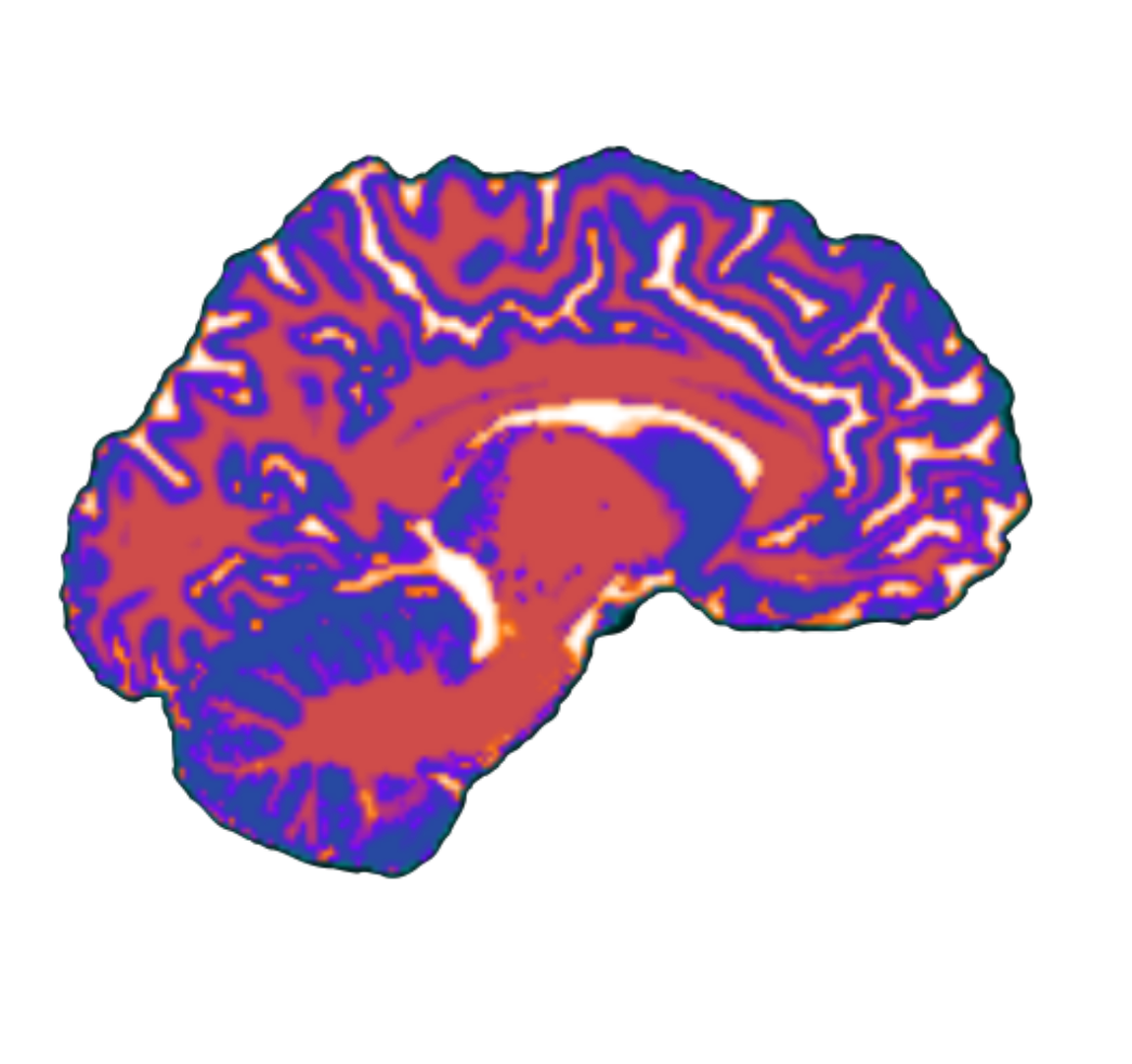
Brain injury in premature infants
Many of these observations come from the following paper: Volpe, 2009
- Cerebellar abnormality is particularly characteristic of premature infants with VLBW.
- Study: 41 premature infants - 25-30% exhibited neuronal loss in the cerebellum
- MRI studies have been particularly valuable in the identification of cerebellar disease in premature infants.
- Cerebellar development
- The cerebellum develops especially rapidly in the last half of human gestation. This result was highlighted by Dobbings et al. (1973; 1974).
- Volumetric MRI study of premature infants has documented an approximately three-times increase in cerebellar volume from 28 to 40 weeks’ gestation.95 Indeed, the rate of overall growth during this period exceeds that of the cerebral cortex, and both neuronal proliferation and migration are prominent ten-Donkelaar et al. (2003; 2006)
- Cerebellar vulnerability
- Rapidly developing process that shows vulnerability.
- This vulnerability of the cerebellum during its phase of rapid growth was documented several decades ago in experimental models of undernutrition, x-irradiation, and glucocorticoid exposure (Dobbings et al. 1970; Dobbings et al. 1974; Chase et al. 1969; Howard, 1968);
- The fundamental nature of cerebellar disturbance in premature infants is unclear. Except for the minority who have cerebellar hemorrhage, overt tissue destruction is absent. The excitatory interaction between the cerebellum and cerebral cortex via corticopontine tracts and then pontocerebellar connections might be crucial for cerebellar development (Limperpaoulos et al. 2005)
- The occurrence of marked cerebellar growth failure in premature infants has almost been completely confined to infants of less than 32 weeks gestation and most commonly of 24-28 weeks’ gestation. Most striking developmental event at this time is the proliferation and inward radial migration of external granule cells is centered on the surface of the cerebellum. The outermost portion of the EGL contains the proliferating cells, exposure of these cells to noxious compounds in the CSF could be deleterious.
- The negative effects of cerebellar growth in the small premature infant could relate to direct effects on the cerebellum and to effects on cerebellar connections. Concerning the latter, the negative effects could relate both to the loss of positive afferent effects from the cerebrum and brainstem cerebellar relay nuclei, and to the development of negative retrograde effects from loss of efferent connections to the thalamus and cerebrum.
